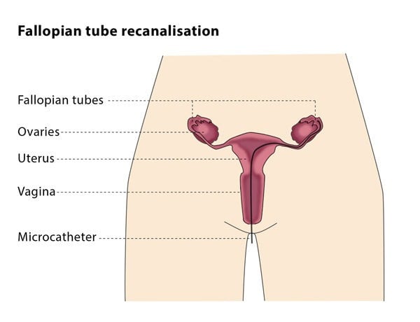
How does the procedure work?
The first part of the procedure is similar to a standard gynaecological exam. The interventional radiologist will insert a speculum into your vagina and will spray a local anaesthetic on your cervix. They will then pass the catheter through your cervix and into your uterus. You may feel some discomfort at this point. The interventional radiologist will inject a few millilitres of a solution that is visible under X-ray through the catheter to make it easier to see the uterus and fallopian tubes during the procedure.
The interventional radiologist will then insert a microguidewire and microcatheter into one of your fallopian tubes and will push or flush the material causing the blockage (often a mucus plug) into your abdominal cavity. The closer to the uterus the blockage is, the more likely it is that the procedure will have the desired outcome. The interventional radiologist will then perform the same technique on your other fallopian tube.
Why perform it?
This procedure can ease infertility that is caused by blockage of the fallopian tubes.
What are the risks?
Tubal perforation (small holes in the fallopian tubes) occurs in about 2% of patients, but this has no severe complications. Pelvic infection is believed to occur in less than 1% of patients. In 3% of patients who get pregnant after the procedure, the embryo is found to be outside the uterus, known as an ectopic pregnancy. It is therefore recommended that you consult your gynaecologist as soon as you know that you are pregnant.

