Cardiovascular and Interventional Radiological Society of Europe
Slider

Nephrostomy

What is percutaneous nephrostomy?

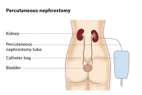
Kidneys are organs in the abdomen which filter the blood and discard the unwanted salts and other products, which are excreted in the form of urine. The kidneys are connected to the urinary bladder (an elastic bag) in the pelvis via tubes called the ureters. Sometimes these tubes get blocked. Due to this blockage, the urine cannot go into the bladder. This creates three problems; one is pain and discomfort which can be felt in the back at the lower end of the ribs transferring all the way in the front near the groin. The second problem is stress on the kidney which causes acute or chronic kidney failure. The third problem is a urinary infection, which can present as fever and chills, or loss of blood pressure if severe.

Obstruction can be caused primarily by three causes. First, a stone in the ureter which causes a block. Second, a tumour (cancer) within the urinary system, either in the ureter, bladder or prostate. Third, a cancer outside the urinary system compressing the ureter.

Solution that can be offered

A double J stent placed by your urologist. Here, the urologist passes a camera into the bladder, visualizes the entry of the ureter into the bladder, and passes a plastic tube that is curved like a J at both ends (hence double J) in under X-ray guidance. One end remains in the kidney, and the other in the bladder, which helps the urine descend into the bladder around the obstruction.

The second option: Percutaneous nephrostomy

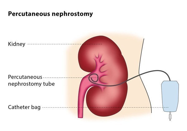
Sometimes, if urologists are not able to pass the tube via the bladder or think it is not a good idea, they recommend a percutaneous nephrostomy. A nephrostomy tube is a thin, flexible tube with resembles a pig’s tail on the inside. The outside looks like a straight plastic tube coming out of the skin in the region of your flank. This tube will be connected to a plastic bag (urine bag). The tube is fixed to the skin with a skin suture. You may also find a butterfly-like dressing holding it in place. You will have a skin dressing over it. Urine leaving the kidney is collected in a plastic bag attached to the tube outside your body. The bag can be strapped to your waist or leg so you can move freely. Once the catheter is in place, you will get pain relief, and if your kidney function is altered, you will notice improvement of the kidney function to how it was before the obstruction happened.

How does the procedure work?

First and foremost, inform your doctor or the team performing the procedures if you are on any anticoagulants (blood thinners) and if you have stopped them or not. Also inform the team if you are allergic to any medications. You will lie on your stomach, usually with one side slightly raised on a pillow or a support. This procedure may be performed under local anaesthesia, local anaesthesia with some intravenous medicine for conscious sedation, or complete general anaesthesia (not frequent). The procedure is performed under sterile conditions. Local anaesthetic will be injected into the skin all the way up to the capsule (protective cover) of the kidney. An incision of 6-8 mm will be made. A needle will be inserted into the kidney under ultrasound guidance. Contrast (X-ray dye) will be injected under X-ray guidance to confirm the position of the needle. Once in position, a metallic wire (guidewire) will be inserted through the needle into the urinary system in the kidney. The needle will be withdrawn over the wire and the catheter will be inserted over the wire into the kidney. Then, once again, dye will be injected to confirm the position of the catheter. Once done, the catheter will be fixed to the skin with a suture.

What are the next steps?

The duration you need the catheter for will be decided by your doctor.

If it is for a stone, it may be kept in place till the infection goes down and stone is removed, or another procedure can be performed.

If it placed for tumour, you may need to keep it in for a longer period or indefinitely. You will need to discuss this with your physician or oncologist.

This catheter will need to be changed every 4-6 weeks. This will be done in the interventional radiology department under Xray guidance.

What are the risks?

Some patients report minor bleeding from the kidney after a nephrostomy. Severe bleeding that requires treatment occurs in less than 5% of patients.

In less than 1/500 patients, an adjacent organ is injured during the nephrostomy.

Although a temporary low-grade fever is common after the procedure, a high fever occurs in around 1-3% of patients, which is usually the result of germs getting into the blood stream after or during the procedure. This can happen if the nephrostomy is being placed into an infected system (if you have had fever with or without chills).